Unveiling the Secrets of the Jumping Spider Anatomy
The body of a jumping spider is a very unique thing. Many people find spiders disturbing, mostly due to their spindly legs and unpredictable movements, but the jumping spider anatomy can be just as off-putting if you don’t understand it.
Two main sections make up parts of a jumping spider; the upper half (the cephalothorax) and the lower half (the abdomen). The eight legs and other appendages of a jumping spider are part of the upper half, with the lower half containing the gut, lungs, heart, reproductive organs, and silk glands. These two segments are joined by the pedicel, which is the spider’s waist.
To better understand the abilities and lives of all jumping spider species, including the regal jumping spider, it is wise to have knowledge of their body parts.
Cephalothorax - The Top Half of the Jumping Spider Anatomy
This segment of the spider combines the head and thorax. The eyes, mouth parts, and legs are the main body parts found on the cephalothorax.
But within the cephalothorax, there are also the muscles, brain ganglia, venom glands, and muscular stomach.
Much like the hard shell of a crab, the top of the cephalothorax is covered by a hard, protective structure called the “carapace.” The underside of this half is covered by the sternum. Both of these structures protect the softer insides of the spider’s body
Eyes
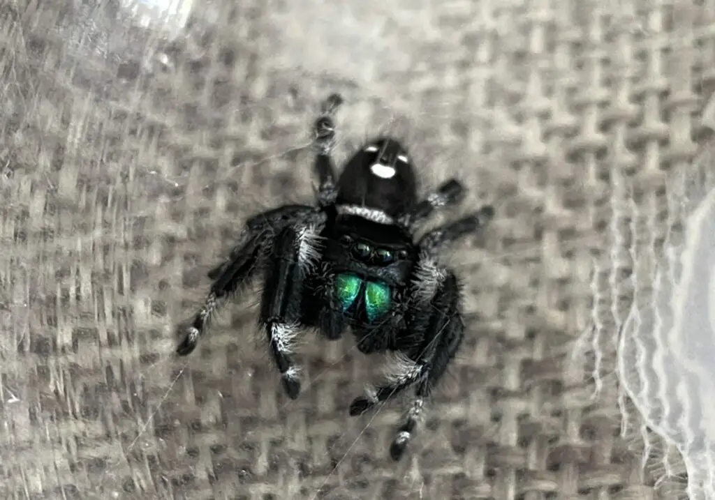
Jumping spider eyes are located on the head. For a jumping spider, like the phidippus audax, there are four pairs of eyes, eight eyes in total, with each pair of eyes decreasing in size and wrapping further around the spider’s head to allow periphery views. They are positioned in three rows. It is this setup that allows spiders to have the best vision.
The first set of eyes, located right at the front of the spider’s head, are the largest. It is this feature that adds to how cute jumping spiders can be; some people will note that the first pair of eyes are almost puppy-like. Not only so they make this species of spider look cute and friendly, but the larger set of eyes is much like our own, as humans. They have high resolution and are stereoscopic, meaning they can detect distances.

The three other pairs of eyes are considered “secondary eyes”. They are much smaller.
Jumping spiders aren’t the only species of spider to have four sets of eyes; this is a common trait for spiders that need to hunt, instead of using webs to catch their prey. It allows them excellent vision to sneak up and capture their prey. The retinas within their tiny eyes are able to rotate, and this allows them to track the movement of their prey; such as ants, flies, and other insects around them. It also helps them stay aware of any predators approaching, such as bigger spiders or animals.
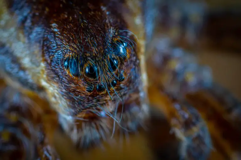
But you might not be aware that different families of spiders have different eye patterns. You can see here the difference between the eye pattern of jumping spiders and that of wolf spiders.
Mouth Parts
Jumping spider mouth anatomy consists of two main parts; the pedipalps and the chelicerae, which are the most visible parts of spiders’ mouths.
Pedipalps
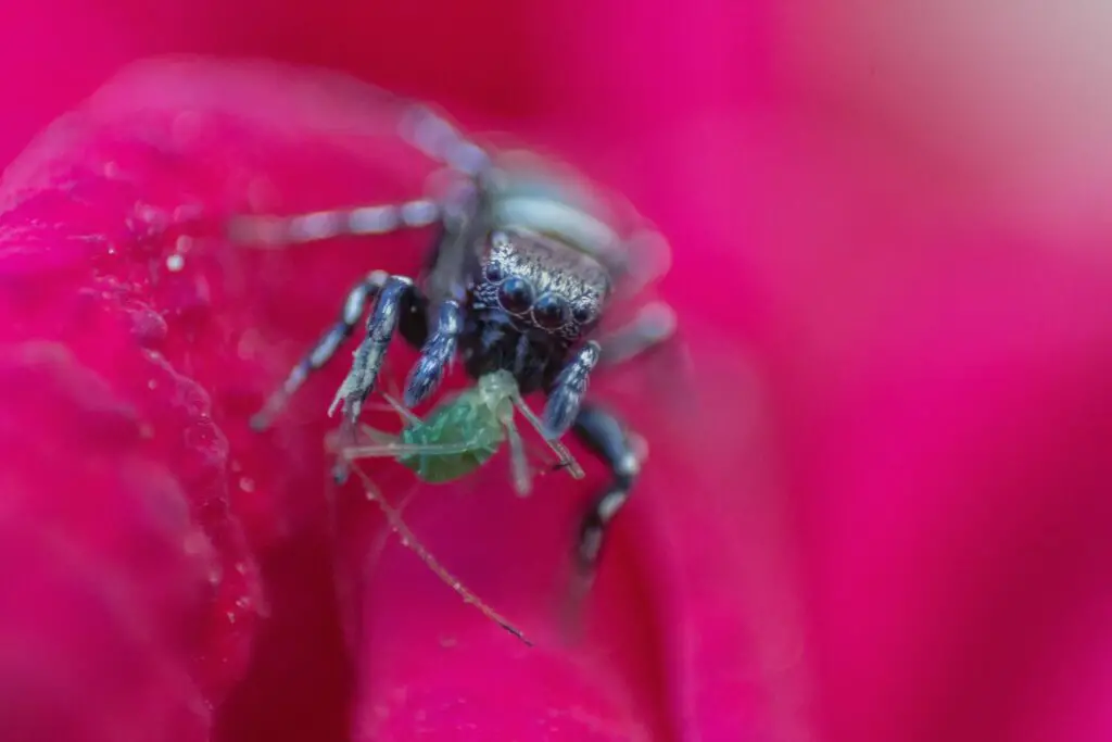
Pedipalps are appendages between the spider’s front legs and chelicerae. It can be easier to think of them like arms, although many people do mistake them for another set of legs. They are often used to catch prey and hold it tight as the spider feeds.
The pedipalps can also be used as an indicator to tell the difference between male and female jumping spiders.
Chelicerae
The chelicerae are the jaws of the spider, and are often tipped with fangs, which is useful when catching insects. Although the chelicerae might not be very strong, the production of venom from the chelicerae can aid them during feeding or protecting themselves. The venom glands are stored in the chelicerae, and may extend into the cephalothorax.
The chelicerae can be a variety of colors; blue, green, pink, gold, etc. They can even change during the molting process that occurs multiple times in a spider’s lifetime.
Although some believe you can check the sex of a spider by the color of chelicerae, this is not a fool-proof method. Some colors are thought to be more common for males, and the same is thought for female spiders, but this is not a guarantee. You would be better to judge the sex of a spider by looking at the pedipalps or epigyne.
Legs
It is common knowledge that most spiders have eight legs, and jumping spiders are no different. The legs contain seven segments; coxa, trochanter, femur, patella, tibia, metatarsus, and tarsus.
However, as their name suggests, this particular species of spider is well-known for its ability to jump. This means a great deal of strength must come from the spider’s legs to ensure it can jump efficiently. The strength of jumping spider legs is unquestionable, especially as some can leap up to 40 times their body length.
Their impressive ability to jump comes from quickly straightening their back legs.

Abdomen - The Bottom Half of the Jumping Spider Anatomy
Another part of the anatomy of jumping spiders is the abdomen. A spider’s abdomen has the ability to expand and contract, depending on the conditions of the spider. This is useful when feeding and also when females are carrying eggs.
Book Lungs
A spider’s lungs can be described similarly to a book – hence the name. The lungs open and close, spreading outwards before closing back in on themselves. They are made up of several thin membranes, almost like pages of a book. Depending on the species, there could be up to 80 membranes making up the spider’s book lungs.
The book lungs, which work in tandem with the tubular tracheae in the spider’s circulatory system, are used to extract oxygen from the air and also to release carbon dioxide.
Spinnerets
This part of the spider’s anatomy is at the very rear of their abdomen. The spinnerets are the silk spinning organs, of which there are usually between four and six.
The spinnerets can be used for a variety of purposes; spinning webs, creating egg sacs, and even creating drag lines that the spider can use to travel, particularly when on vertical surfaces.
Silk Glands
As the name suggests, these glands are responsible for producing the liquid protein that makes a spider’s silk. The silk is produced in the spinnerets, and the amount and quality of the silk can vary depending on the spider.
Reproductive Organs of the Jumping Spider Anatomy
On the underside of the abdomen, mature females have a slight indent; it is almost like a belly button on humans. This, however, is the spider’s epigyne. The epigyne is the female reproductive organ, and it is used to deposit eggs into a web or an egg sac.
The jumping spider anatomy for the male spider is different with its reproductive organ located on the front of the abdomen and is used to inseminate the female.